Machine learning and computer vision have found applications in medical science and, recently, pathology.
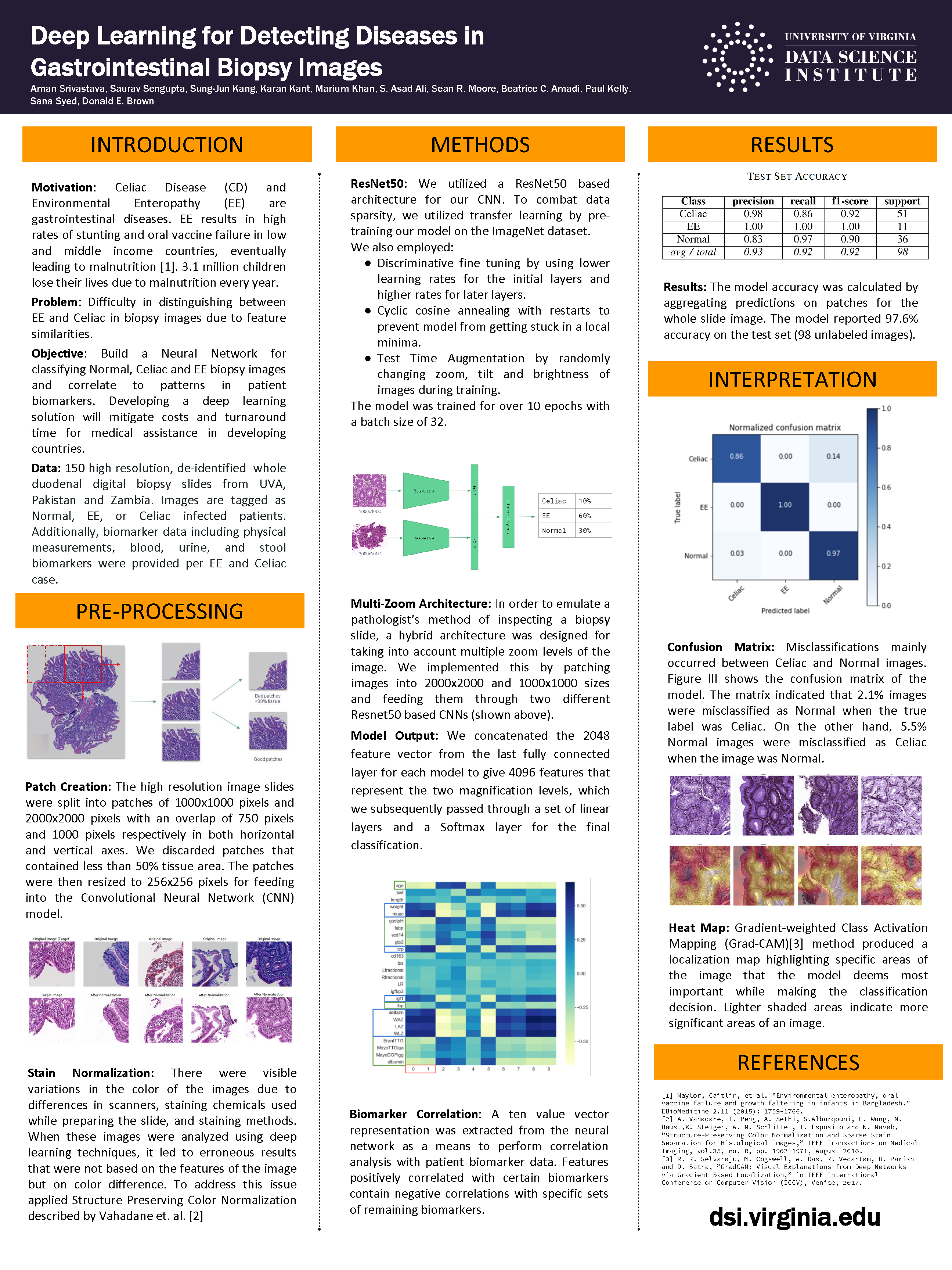
 In particular, deep learning methods for medical diagnostic imaging can reduce delays in diagnosis and give improved accuracy rates over other analysis techniques. Researchers Aman Shrivastava, Saurav Sengupta, Karan Kant and Luke Kang—working with S. Asad Ali (Aga Khan University, Pakistan); Sean Moore (University of Virginia); Beatrice Amadi (University of Zambia, Zambia); Paul Kelly (Queen Mary University of London, United Kingdom); and Sana Syed and Donald Brown (University of Virginia)—undertook a capstone project as part of the Master of Science in Data Science program to investigate methods with applicability to automated diagnosis of images obtained from gastrointestinal biopsies.
These deep learning techniques for biopsy images may help detect distinguishing features in tissues affected by enteropathies. Learning from different areas of an image, or looking for similar patterns in new images, allow for the development of potential classification or clustering models. Techniques like these provide a cutting-edge solution to detecting anomalies.
The research team explored state of the art deep learning architectures used for the visual recognition of natural images and assess their applicability in medical image analysis of digitized human gastrointestinal biopsy slides.
Shrivastava, Sengupta, Kant and Kang were awarded best poster at the 2019 Systems and Information Engineering Design Symposium.

